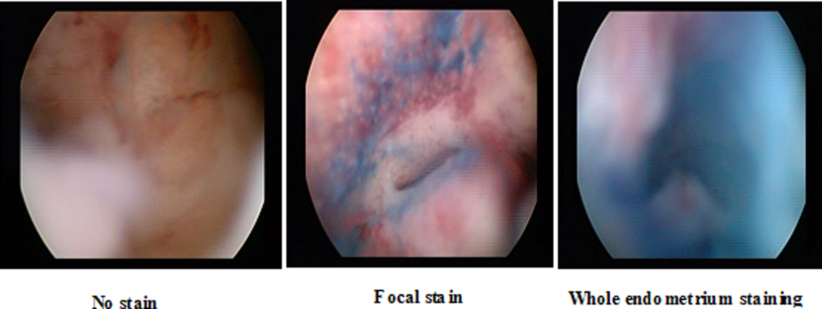
Figure 1. Different patterns of staining at chromohysteroscopy.
| Journal of Clinical Gynecology and Obstetrics, ISSN 1927-1271 print, 1927-128X online, Open Access |
| Article copyright, the authors; Journal compilation copyright, J Clin Gynecol Obstet and Elmer Press Inc |
| Journal website http://www.jcgo.org |
Original Article
Volume 3, Number 1, February 2014, pages 35-41
The Value of Chromohysteroscopy in the Assessment of Postmenopausal Vaginal Bleeding
Figure
Tables
| Number of cases | Mean | SD | Minimum | Maximum | Median | |
|---|---|---|---|---|---|---|
| Age distribution (in years) | 50 | 57.5 | 6.3 | 47 | 72 | 58 |
| Parity | 50 | 3.4 | 1.3 | 1 | 6 | 3 |
| Number | Mean | Std deviation | Median | Min. | Max. | Kruskal-Wallis test | ||
|---|---|---|---|---|---|---|---|---|
| Chi-square | P value | |||||||
| *: Statistically significant. NEP: No endometrial pathology. | ||||||||
| Atrophy | N=14 | 4.4 | 1.2 | 4.0 | 3.0 | 8.0 | 36.611 | < 0.001* |
| Endometritis | N=5 | 8.0 | 3.7 | 6.0 | 5.0 | 12.0 | ||
| Endometrial hyperplasia | N=4 | 16.5 | 4.7 | 15.5 | 12.0 | 23.0 | ||
| Endometrial cancer | N=4 | 25.3 | 5.1 | 25.0 | 20.0 | 31.0 | ||
| NEP | N=23 | 8.0 | 2.2 | 7.0 | 5.0 | 13.0 | ||
| Chromohysteroscopy pathology | Chromohysteroscopy stain | ||||
|---|---|---|---|---|---|
| Focal stain | Whole end | No stain | Total | ||
| NEP: No endometrial pathology. | |||||
| Atrophy | Count | 12 | 0 | 2 | 14 |
| % within chromohyst pathology | 85.7% | 0% | 14.3% | 100.0% | |
| % within chromohys stain | 31.6% | 0% | 28.6% | 28.0% | |
| Endometritis | Count | 5 | 3 | 0 | 8 |
| % within chromohyst pathology | 62.5% | 37.5% | 0.0% | 100.0% | |
| % within chromohys stain | 13.2% | 60.0% | 0.0% | 16.0% | |
| Endo hyperplasia | Count | 6 | 0 | 0 | 6 |
| % within chromohyst pathology | 100.0% | 0.0% | 0.0% | 100.0% | |
| % within chromohys stain | 15.8% | 0.0% | 0.0% | 12.0% | |
| Endo cancer | Count | 2 | 2 | 0 | 4 |
| % within chromohyst pathology | 50.0% | 50.0% | 0.0% | 100.0% | |
| % within chromohys stain | 5.3% | 540.0% | 0.0% | 8.0% | |
| NEP | Count | 13 | 0 | 5 | 18 |
| % within chromohyst pathology | 72.2% | 0.0% | 27.8% | 100.0% | |
| % within Chromohys stain | 34.2% | 0.0% | 71.4% | 36.0% | |
| Total | Count | 38 | 5 | 7 | 50 |
| % within chromohyst pathology | 76.0% | 10.0% | 14.0% | 100.0% | |
| % within chromohys stain | 100.0% | 100.0% | 100.0% | 100.0% | |
| Atrophy | Endometritis | Endo hyperplasia | Endo cancer | NEP | Total | Pearson’s Chi-square | ||
|---|---|---|---|---|---|---|---|---|
| Value | P value | |||||||
| *: Statistically significant. | ||||||||
| Fractional curettage (cases) | 14 | 5 | 4 | 4 | 23 | 50 | 33.016 | < 0.001* |
| Chromo-hysteroscopy (cases) | 14 | 8 | 6 | 4 | 18 | 50 | ||